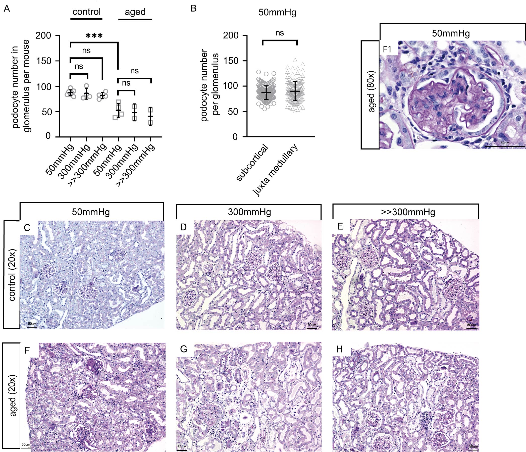
Fig. 2. Podocyte loss in healthy mice. (A) Total podocyte number per mouse at baseline and after high (300 mmHg) or maximal perfusion pressures (>>300 mmHg) (each circle represents 1 kidney (40 glomeruli pro mouse, n=7 control mice, n=4 aged mice), ANOVA, ***P<0.001 and ns = not statistically significant; error bars represent means ± SD). (B) Total podocyte number per glomerulus in subcortical and juxtamedullary glomeruli at baseline (each circle represents 1 subcortical glomerulus and each triangle 1 juxtamedullary glomerulus (n=20 per mouse), ANOVA, ns = not statistically significant; error bars represent means ± SD). (C-E) Histologic staining of young mice with periodic acid-Schiff at baseline and after perfusion with higher pressures (300mmHg and >>300mmHg), showing only mild changes, i.e tubular dilatation and tubulointerstitial edema. (F-H) Histologic staining of aged mice at baseline showing mesangial expansion and occasional sclerosis (arrow, F1) and after perfusion with 300mmHg and supramaximal pressure showing tubulointerstitial dilatation (G-H); ctrl: control mice.